LAMINATE / Single timepoint analysis
A closer look at
one MRI session.
Combine FLAIR and SWI from a single session into a FLAIR* image that makes veins within lesions easier to see, with optional AI lesion segmentation to review.
What you bring
- One MRI session of a subject.
- A FLAIR and an SWI series, as DICOM.
- An optional Phase series, shown as a fourth image.
- An uploaded archive, or studies sent from your PACS through the on-site gateway.
What you get
- A FLAIR* image: the SWI aligned to the FLAIR and combined with it.
- An AI lesion mask, when you switch segmentation on.
- The FLAIR* series as DICOM, stored with the session.
- A report of lesions, volume and your scores, as HTML or PDF.
What happens to a session
Convert and align
The DICOM series are converted and the SWI is registered to the FLAIR.
Build the FLAIR* image
The aligned SWI and FLAIR are combined into one image that makes veins and tissue easier to see.
Segment lesions (optional)
Brain extraction and AI lesion segmentation can be switched on for each analysis. Segmentation runs on a GPU server.
Return the results
The FLAIR* series is added to the same session, and the lesion mask is ready for review.
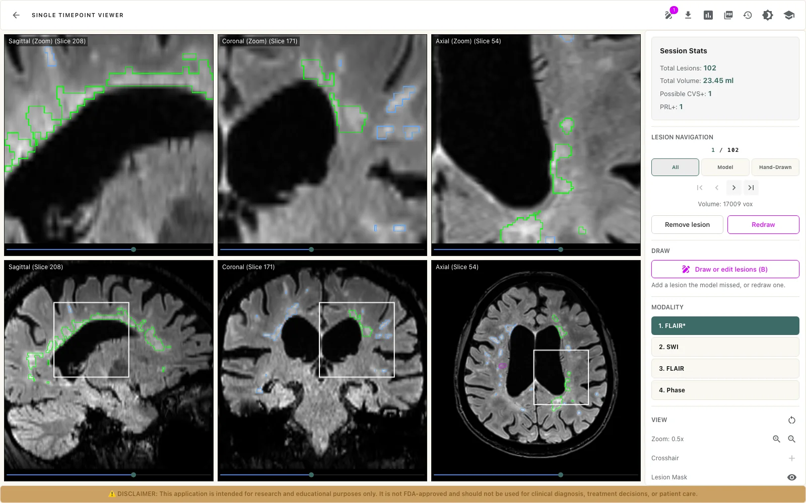
Review in the Single Timepoint Viewer
- Switch between FLAIR*, SWI, FLAIR and Phase, with the slice kept in place.
- Three planes with a crosshair, zoom, and window and level for each image.
- Step through lesions with first, previous, next and last.
- Score each lesion’s likelihood of a central vein, and mark a paramagnetic rim, by hand.
- Remove a lesion, redraw it, or draw one the model missed.
- Totals for the session: lesions, volume, possible central vein sign and paramagnetic rim.
- Edits save automatically, with undo, redo and a version history.
what it does not do
Good to know
- Central vein sign and paramagnetic rim are scored by the reviewer. LAMINATE does not detect them automatically.
- Brain extraction and AI lesion segmentation are options for each analysis. Without segmentation there is no lesion mask to review.
- The FLAIR* series can be sent back to your PACS through the on-site gateway.
This application is intended for research and educational purposes only. It is not FDA-approved and should not be used for clinical diagnosis, treatment decisions, or patient care.