LAMINATE / Longitudinal analysis
See the change
between sessions.
Compare a baseline and a follow-up session of the same subject. LAMINATE labels each lesion as new, growing, shrinking, static or gone, and reports lesion count, volume and the change in lesion load.
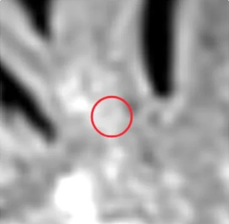
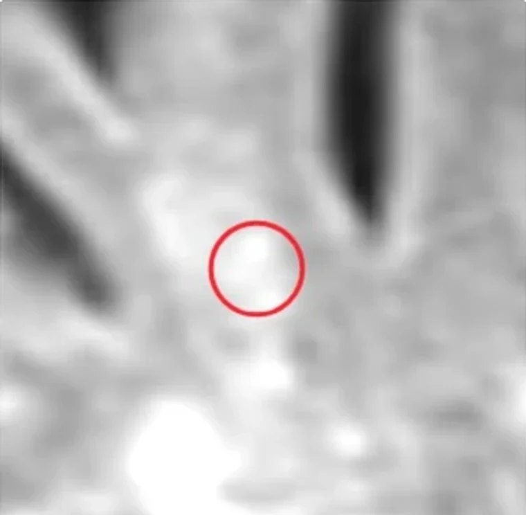 BaselineFollow-up
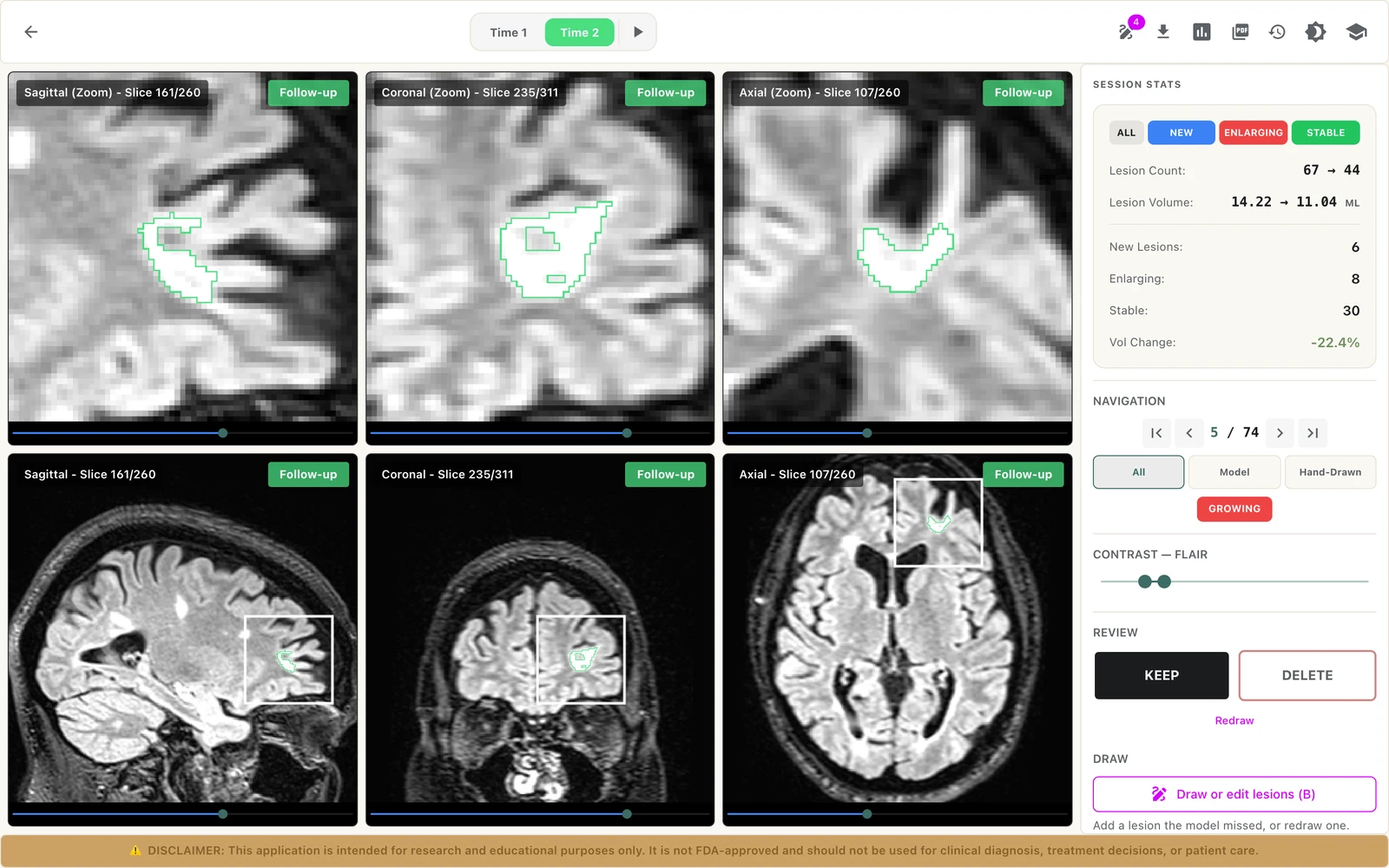
a real screen from LAMINATE
What you bring
- Two sessions of one subject: a baseline and a follow-up.
- A FLAIR and a T1-weighted series in each session, as DICOM.
- An uploaded archive, or both studies sent from your PACS in one upload.
What you get
- Lesion segmentations for both sessions, registered to each other.
- Each lesion labelled New, Growing, Shrinking, Static or Gone.
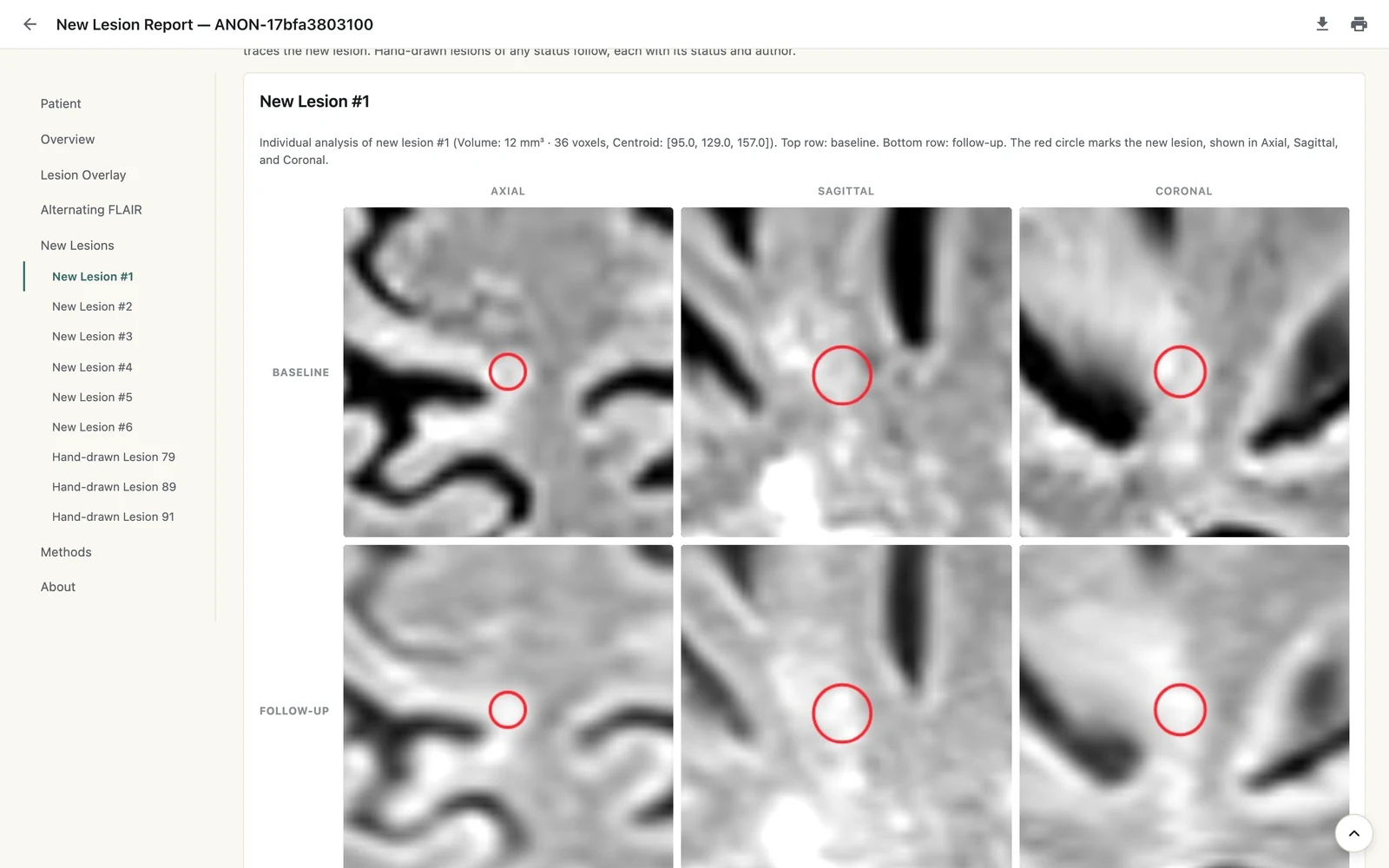
- A report with lesion count, lesion volume, new lesion count and volume, expanding lesion volume and percent change in lesion load.
- Lesion figures, and NIfTI volumes you can download.
What happens to a pair of sessions
Align the sessions
The baseline and follow-up images are registered to each other.
Segment lesions
Lesions are segmented in both sessions.
Match and label
In the viewer, lesions that overlap across the sessions are matched, so merges and splits are handled. Each lesion gets its status from the change in its volume.
Report the change
Measurements and figures are collected into a report you can view, download as HTML or save as PDF.
Review in the Longitudinal Viewer
- Switch between baseline and follow-up with one key, or alternate them automatically.
- Axial, sagittal and coronal views of both sessions, with crosshair, zoom and contrast.
- A lesion list with each lesion’s status, volume and review progress. Jump to any lesion.
- Keep or delete each lesion, which changes what the report counts.
- Add a lesion the model missed, or redraw one. Clone a lesion’s shape between sessions, merge, or reset to the model output.
- Edits save automatically, with undo, redo and a version history.
what it does not do
Good to know
- LAMINATE compares two sessions at a time. Each pair is its own analysis; trends across several visits are not available yet.
- Both sessions must belong to the same subject, with FLAIR and T1-weighted images in each.
- Results are NIfTI volumes, figures and a report. They are not returned to PACS as DICOM today.
- An analysis starts automatically from PACS only when both studies arrive in one upload.
This application is intended for research and educational purposes only. It is not FDA-approved and should not be used for clinical diagnosis, treatment decisions, or patient care.